Pelvic organ prolapse
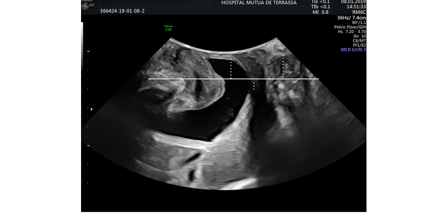
In addition to providing information about the physiopathology of prolapse – as we will see in the next section – ultrasound imaging also allows us to quantify prolapse it in a very simple way. Starting from the sagittal plane, prolapse can be quantified using the pubic symphysis as a line of reference.
Ultrasonographic cut-off points, from which the patients show symptoms, have been described; these can be correlated directly with applicable measurements in the POP-Q system (3). A woman usually shows symptoms when: a cystocele is at least 10 mm below the pubic symphysis (Ba > 0.5); a uterine prolapse is 15 mm above the pubis (C > 5); or a rectocele is at least 15 mm below the pubic symphysis (Bp > - 0.5) (Figure 1).
If we analyse this pathology compartment by compartment, the ultrasound helps us understand the type of prolapse we are dealing with.