Ultrasound in the study of urinary and faecal incontinence

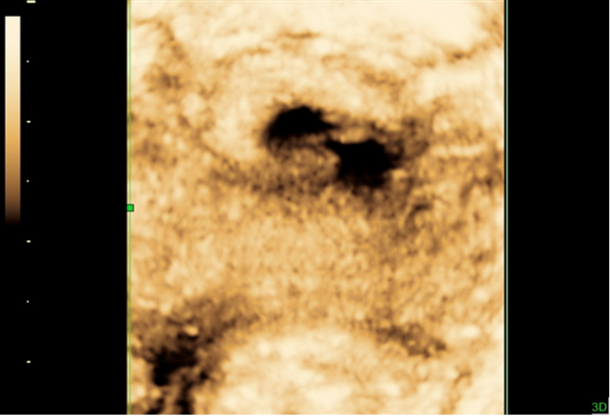
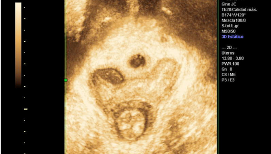
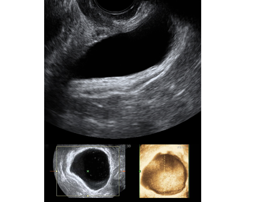
While the three-dimensional image gives us a great deal to work with for examining the levator ani, the two-dimensional image provides us with enough information to make a correct diagnosis of urinary incontinence and conduct a good anatomical and functional study. In this case, the axial plane has very little purpose because the sagittal plane suffices for assessing the main parameters, which consist of both urethral mobility and the sphincter.

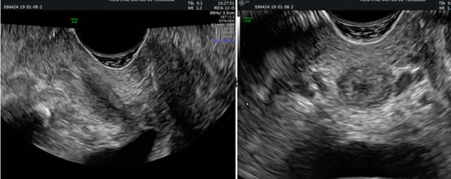
The ultrasound allows us to clearly see the urethra, which is visualized ultrasonographically as either an anechogenic or hypoechogenic central area surrounded by a more echogenic area that comprises the urethral sphincter, which we can see both in the sagittal plane and in the transverse plane (Figure 1). The vascularization of the urethra-sphincter complex can also be assessed by Doppler.

Figure 1. The urethra in the sagittal plane (on the left) and in the transverse plane (on the right).
Injuries such as urethral diverticulum can be easily observed in both two-dimensional and three-dimensional ultrasounds as anechogenic or hypoechogenic saccular structures that connect with the urethral lumen and that do not capture Doppler signals (Figure 2). In turn, we can differentiate between pathologies near the urethra that can sometimes look like a urethral diverticulum, such as a Gartner cyst (Figure 3). These cystic images are usually perfectly individualized and do not come into contact with the urethral lumen.
Figure 2. Urethral diverticulum
Figure 3. Gartner cyst
Next, we examine the entire surface of the bladder in order to rule out any exophytic lesions. Both two and three-dimensional ultrasounds can clearly depict the inside of the bladder (Figure 4). The three-dimensional images are very similar to what we would see in a cystoscopy.
Figure 4. Two-dimensional (in greyscale) and three-dimensional (in colour) images of the inside of the bladder
Doppler technology can help us in these cases, since neoformative processes in the bladder are usually hypervascular. Given that the Doppler is a dynamic test, we're also able to see the ejections from the ureteral meatus into the urinary bladder (Clip 1). This may be useful in some types of surgery in order to check ureteral indemnity.
Other interesting topics:
Post-void residual test - Read more

Stress urinary incontinence - Read more

Overactive bladder - Read more

Faecal incontinence - Read more

Pelvic floor rehabilitation - Read more

Bibliography - Read more