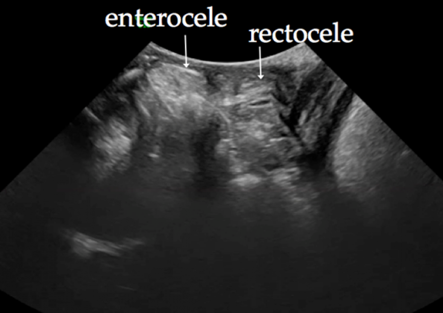
Ultrasound can help us the most in cases of prolapse of the posterior compartment because it identifies the prolapsed organ – whether a rectocele (Clip 2), an enterocele or a combination of both (Figure 3 and Clip 3).
Figure 3. The image shows a posterior compartment prolapse in which we can clearly see a rectoenterocele
The rectocele behaves like the cystocele: that is, when the rectocele extends beyond the anal canal, there is increased voiding dysfunction or obstructive defecation because the rectovaginal septum is torn. On the other hand, when the rectovaginal septum is intact, there is no obstructive defecation; what occurrs instead is perineal descent (3).
In many cases, a clinical examination only allows us to identify a prolapse of the posterior compartment, but it does not allow us to distinguish exactly what is involved. The ultrasound can clearly differentiate between a rectocele, an associated enterocele, or a pure enterocele. This is an important difference to determine, given that the surgical treatment for each of these entities is different.